Plantar fasciitis is one of the most common causes of heel pain worldwide, accounting for roughly 10% of all running-related injuries and millions of clinical visits annually. Characterized by sharp, localized pain at the anteromedial aspect of the calcaneus, the condition stems from microtears and chronic degeneration of the plantar fascia—a thick, fibrous band of connective tissue spanning the longitudinal arch of the foot. The hallmark symptom is debilitating pain during the first few steps in the morning or after prolonged periods of rest, a phenomenon directly linked to nocturnal foot positioning.
While conservative treatments range from non-steroidal anti-inflammatory drugs (NSAIDs) and corticosteroid injections to custom orthotics and physical therapy, night splints have emerged as a cornerstone of mechanical intervention. By holding the foot in a constant state of dorsiflexion during sleep, night splints intercept the pathological cycle of healing and re-tearing that defines the condition.
Pathophysiology and the “First-Step” Phenomenon
To understand how night splints function, one must first examine the anatomical structure they are designed to treat. The plantar fascia originates at the medial tubercle of the calcaneus and inserts into the proximal phalanges of the toes. It functions as a dynamic shock absorber and structural tie-rod, supporting the medial longitudinal arch via the windlass mechanism. During gait, as the toes dorsiflex, the plantar fascia winds around the metatarsophalangeal joints, shortening the band and elevating the arch to provide a rigid lever for propulsion.
When subjected to repetitive overload, structural microtrauma occurs at the calcaneal insertion. The body attempts to repair these microscopic tears through a fibroblastic response. However, when an individual sleeps or rests, the foot naturally drops into a position of plantar flexion (the toes point downward), accelerated by the weight of bed coverings and the natural contraction of the gastrocnemius and soleus muscles. In this plantar-flexed state, the plantar fascia shortens and adapts to a contracted length. As the fibroblastic repair occurs overnight, the tissue mends in this shortened configuration.
The crisis occurs upon waking. The moment the individual steps out of bed, body weight forces the foot into a neutral or dorsiflexed position, violently stretching the newly mended, shortened fibrous tissue. This sudden mechanical stress causes acute micro-tearing of the healing fascia, triggering the intense, sharp pain characteristic of the “first-step” phenomenon. This daily cycle of nocturnal shortening followed by morning tearing creates a chronic, degenerative cycle (plantar fasciosis) rather than an acute inflammatory state, severely delaying long-term recovery.
Biomechanical Mechanisms of Night Splints
Night splints are designed specifically to break this cycle by altering nocturnal foot geometry. By enforcing a consistent angle of neutral to slight dorsiflexion (typically between 90 degrees and 95 degrees relative to the lower leg), the splint maintains a continuous, low-load passive stretch on both the plantar fascia and the Achilles tendon complex throughout the sleep cycle.
This prolonged static stretching achieves several critical physiological outcomes:
- Prevention of Nocturnal Contracture: By maintaining the structural length of the fascia overnight, the body’s natural tissue repair processes occur while the ligament is extended. Consequently, the first morning steps do not induce the sudden, violent elongation that ruptures new collagen fibers.
- Mitigation of the Windlass Trigger: Keeping the foot at a right angle prevents the toes from dropping, minimizing the baseline tension spike that occurs when transitioning to weight-bearing activities.
- Reduction of Gastrocnemius-Soleus Tightness: The calf muscle complex shares a continuous structural connection with the plantar fascia through the paratenon of the Achilles tendon at the calcaneus. A tight calf muscle accelerates the pulling force on the heel bone, exacerbating fascial strain. Sustained nocturnal dorsiflexion lengthens these posterior structures, decreasing the aggregate tensile load experienced by the calcaneal insertion during daytime ambulation.
Clinical Efficacy and Comparative Research
Extensive clinical research supports using night splints as part of a multimodal treatment strategy. Multiple randomized controlled trials and systematic reviews indicate that night splints significantly reduce visual analog scale (VAS) pain scores, particularly for morning symptoms, and accelerate overall recovery times compared to isolated treatments.
Clinical studies demonstrate that while night splints are highly effective, they achieve optimal outcomes when paired with specific complementary interventions:
| Intervention Combination | Clinical Efficacy Summary | Primary Therapeutic Value |
| Night Splint + Eccentric Calf Stretching | Exceptionally high success rates; fastest reduction in morning VAS scores. | Addresses both nocturnal contracture and daytime kinetic chain tightness. |
| Night Splint + Custom Orthotics | Highly effective for long-term functional correction and pain maintenance. | Splint manages resting length; orthotics manage dynamic alignment during weight-bearing hours. |
| Night Splint Alone | Moderate efficacy; resolves symptoms but over a prolonged timeline. | Prevents morning re-injury but lacks dynamic daytime structural support. |
Research underlines that night splints do not necessarily alter the structural thickness of a chronically degenerated fascia, but they dramatically alter tissue tolerance to mechanical load. By eliminating the daily trauma of the morning’s first steps, they create a stabilized physiological environment that allows secondary therapies—like physical therapy or shockwave treatment—to work more efficiently.
Design Typologies: Posterior vs. Anterior Splints
Orthopedic manufacturers generally design night splints in two primary configurations, each presenting distinct mechanical and user-experience profiles:
1. Posterior Boot Splints
The traditional posterior splint features a rigid plastic shell running down the back of the calf and under the sole of the foot, holding the ankle fixed with adjustable dorsiflexion straps.
- Advantages: Provides rigid, uncompromising immobilization and precise control over the angle of flexion. It is highly effective for individuals with concurrent, severe Achilles tendon tightness.
- Disadvantages: Extremely bulky, heavy, and heat-retaining. It restricts mobility entirely, making nighttime bathroom visits cumbersome and often disrupting sleep patterns.
2. Anterior (Dorsal) Splints
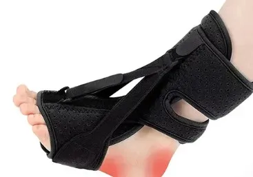
Anterior splints feature a rigid support structure running along the front of the shin and the top of the foot, leaving the heel and plantar aspect exposed.
- Advantages: Lighter, less bulky, and cooler than posterior boots. They allow for safer, more natural ambulation during brief nighttime awakenings.
- Disadvantages: Because the rigid structure sits on top of the foot, it relies heavily on securing straps around the toes and ankle to maintain dorsiflexion. This can sometimes lead to localized pressure points or slight slippage, slightly reducing the absolute rigidity of the stretch.
The Challenge of Patient Compliance
Despite their clear biomechanical utility, the primary limitation of night splints is patient compliance. The therapeutic benefit of a night splint is entirely dependent on wearing time; keeping the device on for less than 4 to 5 hours a night yields negligible results. However, many patients find sleeping in a rigid orthopedic device highly uncomfortable, leading to early removal during the night or total abandonment of the therapy.
To optimize compliance and therapeutic success, clinicians focus on specific patient education and adjustment strategies:
Gradual Accommodation Protocol: Patients should avoid attempting to wear a night splint for a full eight-hour sleep cycle on the first night. Instead, they should wear the device for 1 to 2 hours during evening rest (e.g., while reading or watching television) to desensitize the neurological stretch reflex. The wear time can then be gradually increased over two weeks.
Furthermore, proper strap adjustment is vital. Patients often overtighten the straps in an attempt to maximize the stretch, which compresses local superficial nerves and blood vessels, causing paresthesia (numbness and tingling) or throbbing pain that wakes them up. The device should be set to maintain a neutral 90-degree angle initially, only increasing to active dorsiflexion once tolerance is established.
Night splints represent a highly logical, biomechanically sound intervention for managing plantar fasciitis. By addressing the root cause of the “first-step” phenomenon—the nocturnal shortening and subsequent morning tearing of fascial tissue—they successfully disrupt the chronic degenerative cycle of the condition. While their bulkiness and potential to disrupt sleep present real compliance hurdles, proper device selection, realistic accommodation protocols, and a comprehensive understanding of the underlying mechanics can mitigate these challenges. When integrated into a comprehensive rehabilitation framework that includes targeted stretching and dynamic foot support, night splints remain an invaluable tool for restoring pain-free mobility.